Politica de securitate (editeaza cu modulul Reasigurare pentru clienti)
 Politica de livrare (editeaza cu modulul Reasigurare pentru clienti)
 Politica de returnare (editeaza cu modulul Reasigurare pentru clienti)
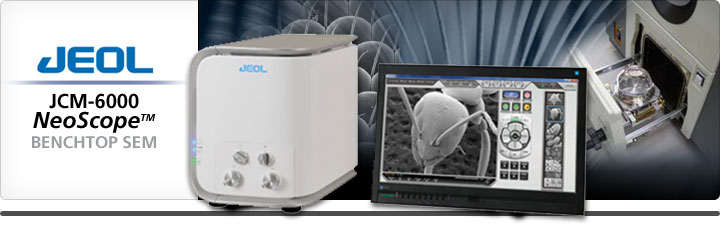
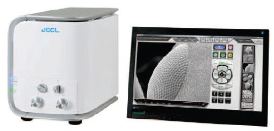
Scanning electron microscope - a powerful yet economic benchtop version from Nikon and JEOL.
JCM-6000Plus
The latest in benchtop SEM technology, the JCM-6000Plus "NeoScopeTM," is a touch panel controlled, multi functional desktop scanning microscope that answers the increasingly diversified needs among users worldwide. The JCM-6000Plus is equipped with the high-sensitivity semiconductor detectors found in high-end instruments, making it easy to acquire composition contrast information about the specimen, and enabling efficient analysis. The series continues to include the high-vacuum functionality and secondary electron detector, offering the ability to clearly observe fine structures on the specimen surface at high magnification.
Main features of the JEOL JCM-6000plus
Automatic image formation after sample introduction within 3 minutes
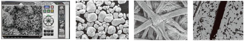
High resolution (60,000X) and large depth of field
Multi-touch screen interface for intuitive operation
Advance automatic functions (focus, stigmation, brightness/contrast)
High and low vacuum modes
Three selectable accelerating voltages
Secondary electron and solid state backscattered electron detector
Large sample coverage (up to 70 mm diameter)
Options include: motor drive stage and EDS
High performance system in a compact, innovative model
Intuitive touch panel operation with new GUI
Well focused high resolution morphological observation
Secondary electron as well as Backscattered electron imaging for compositional distribution
Selectable accelerating voltages
High and Low vacuum operation
Full-featured Energy-dispersive X-ray Spectroscopy (EDS) with SDD technology (Optional)
Metrology supported
Imaging of tilted, rotated samples (Optional)
Touch panel
Ease of operation through the multi-touch screen or standard keyboard/mouse
Compact, light, and energy saving
Compact body JCM-6000
Compact body equal to an optical microscope
Base unit: 330mm (W) x 490mm (D) x 430mm (H); 50 kg
Utility: Single phase 100 V to 240 V, 50/60 Hz, 700 to 960 VA
New capabilities for imaging
Secondary electron imaging and backscattered electron imaging supported at high vacuum
New high sensitivity solid state backscatter electron detector provides both composition and topographic imaging information
Dual frame imaging to facilitate comparison of live and retrieved images
A wide magnification range from the lowest 10x for wide area of view up to 60,000x
Dual frame display
Simultaneous display of live and retrieved images allows for comparative observation
Enhanced low vacuum capability
New solid state backscattered electron detector
Easy observation of non conductive samples in the direct low vacuum mode
Only 2 minutes 30 seconds from sample loading to imaging
Simple operation
Easy touch panel operation
A complete range of automated functions (auto focus, auto stigmator, auto contrast/brightness)
Easy, dependable auto gun alignment (filament centering)
Tilt/rotation motor drive holder
Tilt/rotation motor drive holder
The tilt/rotation motor drive specimen holder allows the operator to tilt and rotate the sample for well focused 3D morphological observation.
Optional accessories
Energy dispersive X-ray spectrometer
Energy dispersive X-ray spectrometer (EDS) for elemental analysis
JEOL's proprietary EDS
Quick, reliable customer support guarantees satisfaction
*This option is retrofittable
| Magnification: | Secondary electron image : × 10 to × 60,000 Backscattered electron image : × 10 to × 30,000 (when image size is 128 mm × 96 mm) |
|---|---|
| Imaging mode: | Secondary electron image, Backscattered electron image (composition, topographic or stereoscopic image) |
| Accelerating voltages: | Secondary electron image ; 5 kV, 10 kV, 15 kV (3 stages) Backscattered electron image ; 10 kV, 15 kV (2 stages) |
| Electron gun: | Small gun with cartridge filament integrating wehnelt |
| Bias current: | Auto bias (linked to accelerating voltage and filament current) |
| Condenser lens: | Two stage electromagnetic zoom condenser lens |
| Objective lens: | Electromagnetic lens |
| Auto magnification correction: | Magnification corrected with reference to sample height (7 mm, WD56 to 53 mm, WD10) |
| Preset magnification: | 6 levels, user programmable |
| Specimen stage: | Manual control for X and Y X: 35mm, Y: 35mm |
| Maximum sample size: | Diameter 70 mm, height 50 mm |
| Specimen exchange: | Draw-out mechanism |
| Image memory: | One, 1,280 × 960 × 16 bits |
| Pixels: | 640 × 480, 1,280 × 960 |
| Image processing: | Pixel accumulation Image accumulation (recursible) |
| Automated functions: | Full-auto, filament adjustment, alignment, focus, stigmator, exposure |
| Metrology: | Distance between 2 points, angles |
| File format: | BMP, TIFF, JPEG |
| Computer: | PC (desktop PC), OS Windows®7 |
| Monitor: | 23 inch wide LCD monitor (touch panel) |
| Evacuation system: | Fully automatic, TMP : 1, RP : 1 |
| Power supply: | Voltage : Single phase AC 100 V (120 V, 220 V, 240 V) 50/60 Hz, 700 VA (AC 100 V) 840 VA (AC 120 V) 880 VA (AC 220 V) 960 VA (AC 240 V) Fluctuation ±10 % or less, with grounding |
|---|---|
| Installation Room: | Room temperature: 15 to 30°C Humidity: 60% or less Operation table: Sturdy table with a loading capacity of 100 kg or more |
| Weight: | Main console : approximately 50 kg RP: approximately 9 kg Power supply box : approximately 10 kg |
| Base unit dimensions: | 325mm (Width) x 490mm (Depth) x x430mm (Height) |